
News & Events
Diastolic Function Made Simple: The ASE 2025 Approach
Diastolic function can be assessed in two simple steps: first confirm that dysfunction is present, then decide whether left atrial pressure (LAP) is high. The 2025 update from the American Society of Echocardiography (ASE) built its algorithm around exactly this logic.
The older 2016 approach left many patients labelled “indeterminate”. The 2025 guideline adds LA reservoir strain (LARS), moves LA volume to a supporting role, and gives a clearer path to a grade. This guide walks through it the way you would at the echo machine.
Step 1: Is e′ reduced?
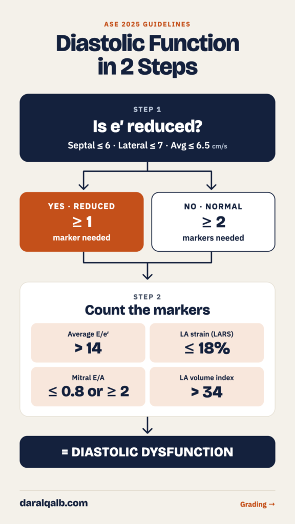
Start with mitral annular e′ by tissue Doppler. It is the most feasible and reproducible marker of LV relaxation, and it is less load dependent than mitral inflow.
e′ is reduced when any of these is met:
- Septal e′ ≤ 6 cm/s
- Lateral e′ ≤ 7 cm/s
- Average e′ ≤ 6.5 cm/s
In younger patients you can use age-adjusted cut-offs instead:
| Age | Septal e′ (cm/s) | Lateral e′ (cm/s) | Average e′ (cm/s) |
|---|---|---|---|
| 20–39 y | < 7 | < 10 | < 9 |
| 40–65 y | < 6 | < 8 | < 7 |
| > 65 y | < 6 | < 7 | < 6.5 |
Sample both septal and lateral sites. e′ is less reliable with significant mitral annular calcification, prosthetic mitral valves, regional wall motion abnormalities at the sampled site, and pericardial disease.
Step 2: Count the markers
Next, count how many of these four markers of LA/LV remodeling and raised filling pressure are abnormal:
- Average E/e′ > 14
- LA reservoir strain (LARS) ≤ 18%
- Mitral E/A ≤ 0.8 or ≥ 2
- LA volume index (LAVi) > 34 mL/m²
The rule is simple:
- e′ reduced + at least 1 marker = diastolic dysfunction
- e′ normal + at least 2 markers = diastolic dysfunction
Before counting LAVi, exclude other causes of LA enlargement: athletes, anemia, atrial fibrillation or flutter, and mitral valve disease. An increased LV mass index (> 95 g/m² in women, > 115 g/m² in men) also supports the diagnosis.
Grading: is LA pressure high?
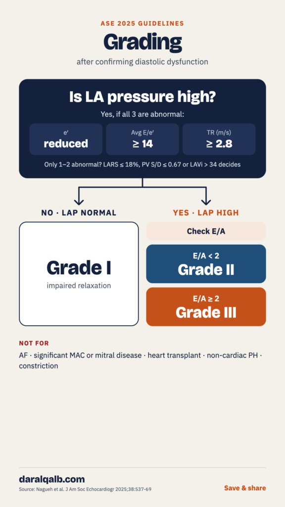
Once dysfunction is confirmed, the grade depends on one question: is LAP normal or high?
LAP is high when all three of these are abnormal:
- Reduced e′ (septal ≤ 6, lateral ≤ 7 or average ≤ 6.5 cm/s)
- Raised E/e′ (average ≥ 14, septal ≥ 15 or lateral ≥ 13)
- Peak TR velocity ≥ 2.8 m/s, or PASP ≥ 35 mmHg
If only one or two are abnormal, use a tie-breaker. LARS ≤ 18%, pulmonary vein S/D ratio ≤ 0.67 or LAVi > 34 mL/m² means LAP is high. IVRT ≤ 70 ms is an alternative.
Then grade:
| Grade | LA pressure | Mitral E/A |
|---|---|---|
| Grade I | Normal | ≤ 0.8 |
| Grade II | High (mild to moderate) | < 2 |
| Grade III | High (marked) | ≥ 2 |
For a patient with exertional dyspnea and Grade I at rest, do not stop there. A diastolic exercise echo is positive when average E/e′ is ≥ 14 (or septal ≥ 15) and TR velocity is > 3.2 m/s.
Why E/A of 2 separates Grade II from Grade III
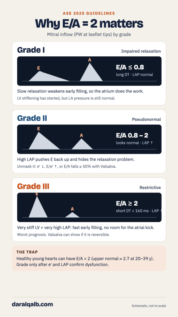
The cut-off of 2 marks the point where the left atrium has almost nothing left to contribute. To see why, remember what the two waves mean. The E wave is the early LA-to-LV gradient; it rises with LAP and falls with slow relaxation. The A wave is atrial contraction, and it depends on how compliant the LV still is in late diastole.
Grade I (impaired relaxation): slow relaxation weakens early filling, so E is small. The atrium compensates, so A is tall. E/A is ≤ 0.8 with a long deceleration time, and LAP is still normal.
Grade II (pseudonormal): relaxation is still impaired, but the raised LAP pushes E back up. The pattern looks normal, with E/A between 0.8 and 2. The reduced e′, high E/e′, or a fall in E/A of 50% or more with Valsalva unmasks it.
Grade III (restrictive): the LV is very stiff and LAP is very high. The early gradient is large, so E is tall, and pressures equalise quickly, so the deceleration time is short (< 160 ms). By late diastole the ventricle is already full and its pressure high, so the atrium contracts against a wall and A is tiny. The result is E/A ≥ 2.
This matters for prognosis. A restrictive pattern carries the worst outlook in dilated cardiomyopathy and HFrEF, and it predicts heart failure hospitalisation in hypertrophic cardiomyopathy. The guideline notes that Valsalva can also separate reversible from irreversible Grade III.
Common pitfalls
Never grade from E/A alone. Healthy young hearts often have E/A above 2; the upper normal limit is about 2.7 at age 20–39. E/A ≥ 2 means restrictive filling only after e′ and LAP confirm dysfunction.
Don’t rely on a single number. A single normal value does not prove normal diastolic function. Look for consistency across the indices, and discard poor-quality signals.
Know where the main algorithm does not apply. Use the dedicated approaches in the guideline for:
- Atrial fibrillation
- Moderate or severe mitral annular calcification
- Any degree of mitral stenosis, or severe primary mitral regurgitation
- Mitral repair, mitral replacement or transcatheter edge-to-edge repair
- Heart transplant recipients
- Non-cardiac pulmonary hypertension
- Pericardial constriction
- LV assist devices
The guideline also does not apply to children, normal pregnancy, or intraoperative studies.
In one line
Confirm dysfunction with e′ and the markers, then ask whether LAP is high: normal means Grade I; high with E/A below 2 is Grade II; high with E/A of 2 or more is Grade III.
Reference
Nagueh SF, Sanborn DY, Oh JK, et al. Recommendations for the Evaluation of Left Ventricular Diastolic Function by Echocardiography and for Heart Failure With Preserved Ejection Fraction Diagnosis: An Update From the American Society of Echocardiography. J Am Soc Echocardiogr 2025;38:537-69. doi:10.1016/j.echo.2025.03.011
This article is for medical education and does not replace clinical judgement or the full guideline.
Very simple way to understand
Excellent explanation
Continue with other cardiology topics
Even write a book !
Best regards
Excellent