Unusual presentation! devil in details
Our patient, female 50 years old with no previous significant medical history.
Two days before the presentation show was waked at night with unbearable epigastric pain and vomiting, so she went to the ER, ER she received IV PPI and referred for ECG
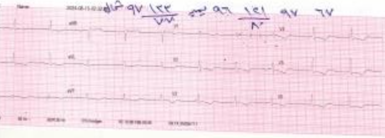
Her ECG at the ER shows diffuse T wave inversion, and her cardiac enzymes were negative:
She was discharged on PPI and was asked to do Echocardiogram later on; her ECHO was done and showed a strange shadow at the free wall of the right atrium, as shown in the video:


What is your opinion and what to do next?
Please leave your comment below
The first complete answer from Dr Ahmed Hamoda
This is a typical case of constrictive pericarditis, proven with 2 main echo signs:
1- septal bounce
2- respiratory variation across the mitral and tricuspid flow
this was proven in the Cath lab while CA, as you can see in the following video the heavily calcified pericardium.